


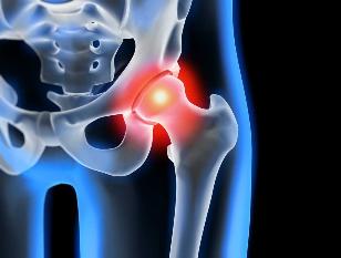
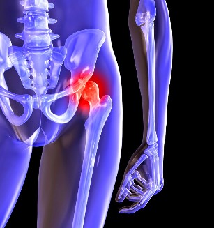
Osteoarthritis of the hip joint degenerative pathology, which is characterized by destruction of üvegporc. The disease develops gradually, associated with pain, reduced range of motion. In the absence of medical intervention at the initial stage of osteoarthritis several years later, there is atrophy of the thigh muscles. The injured limb is shortened, the fusion of the joint space lead to partial or complete paralysis of the hip joint. Cause pathological changes to previous trauma, scoliosis, systemic diseases of musculoskeletal system.
Osteoarthritis is usually diagnosed, the middle-aged and older people. The diagnosis is made up, based on the results of the instrument tests, x-rays, MRI, CT, arthroscopy. Treatment of disease 1 to 2 degrees of severity, conservative. Upon detection of ankylosis, and the ineffectiveness of drug therapy, surgery (arthrodesis, arthroplasty).
The mechanism of development of pathology
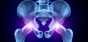
The hip joint consists of two bones — the hip femoral. The lower part of the Basin presents the organization, which is involved in the plexus of the femur, which is the upper part of the acetabulum. During transport, the stationary glenoid fossa and the femoral head move freely. This is the "hinge" device, the hip joint allows you to bend down, stretch, rotate, contributes to the attention to the hips. The smooth gliding of the articular structures provides a smooth, flexible, üvegporc lining of the socket and the femoral head. The main task of the redistribution of the load, when driving, to prevent the rapid deterioration of the bone tissue.
Under the influence of the external or internal factors, nutritional cartilage. There is no circulatory system — nutrients tissue provides the synovial fluid. In the case of arthrosis thickens, it becomes viscous. The deficiency of nutrients provokes the drying out of the surface of üvegporc. This applies to cracks, which leads to a constant micro trauma to the tissue flexion or extension of the hip joint. The cartilage tissue to become thinner, lose their cushioning properties. If you "adapt" to the increase in pressure, the bones become deformed. But in the background, the deterioration of the metabolism of the tissues of the progress of the destructive-degenerative changes.
Causes and predisposing factors
Idiopathic, or primary arthritis develops without a reason. It is believed that the destruction of the cartilage occurs due to the natural aging of the body, slowing down the regenerative processes, the breakdown of collagen and other compounds necessary for the proper regeneration of the structures of the hip joint. Secondary osteoarthritis occurs in the background already present in the body in a pathological condition. The most common causes of secondary disease are the following:
- previous damage — damage-the-tendon unit, muscle tears, to the complete separation of the bone base fractures, sprains;
- malformations of the joints, congenital dysplastic disorders;
- autoimmune disease — rheumatoid, reactive arthritis, psoriatic arthritis, systemic lupus erythematosus;
- non-specific inflammatory diseases, such as suppurative arthritis;
- the specific infections — gonorrhoea, syphilis, brucellosis, anaplasmosis, trichomoniasis, tuberculosis, osteomyelitis, encephalitis;
- disorder in the functioning of the endocrine system;
- degenerative pathology — osteochondropathy of the femoral head;
- hypermobility of the joints due to the production of "super pull" collagen is the trigger of the excessive mobility, weakness, ribbons.
Since the cause of development of osteoarthritis may be hemarthrosis (bleeding in the cavity of the hip joint), the trigger factors include, a violation of hematopoiesis. A prerequisite for the disease of the overweight, excessive exercise, sedentary lifestyle. The development leading to a bad training organisation, the deficiency in dietary products with high content of minerals, fat-and water-soluble vitamins. Postoperative arthrosis occurs a few years after the surgery, especially if it was accompanied by the excision of large amounts of tissue. Osteoarthritis of the hip joint is not transferable by inheritance. But the presence of certain congenital features (violation of metabolism, the structure of the skeleton), the probability that the development will be significantly increased.
Symptoms
Leading symptoms of osteoarthritis of the hip joint pain while walking, the thigh, the knee joint. A person suffering from stiffness in the limbs, stiffness, especially in the morning. Stabilize the joint, the patient begins to limp, my gait has also changed. Over time due to muscle atrophy and deformation of the joint of the limb to be significantly shorter. Another characteristic of the pathology — limitation of hip abduction. For example, difficulties arise when you want to sit in a chair, legs apart on the page.
Even "launched" ARTHRITIS can be cured at home. Just remember, once a day, to smear it...
The arthrosis in the first degree characterized by periodic pain that occurs after intensive physical loads. These are located in the area of articulation disappears after a long vacation.
The second degree osteoarthritis of the hip joint pain intensity increases. Discomfort can also occur the other, extend the hip report, worse when lifting weights or increasing physical activity. Address hip pain, a person begins with a barely noticeable limp. Marked limitation of motion of the joint, especially abduction and internal rotation of the hip.
Arthrosis of the third degree of characterized by constant, severe pain. If you are driving hard, that's why when he walks, a man forced to use a cane or crutches. Since the weak abduction muscles of the hips, balancing the pelvis knot. To compensate for the resulting shortening of the legs of the patient when the movement is tilted toward the affected limb. This provokes the displacement of the center of gravity and increased stress on the joint. At this stage of the arthrosis develops a pronounced ankylosis of the joint.
| Degree | Radiological signs of |
| First | Changes are expressed not sharply. The joint gap moderately, irregularly constricted, that there is no destruction of the surface of the femur. The outer or the inner edge of the acetabulum, there was a small bony lumps |
| Second | The height of the joint space is significantly reduced, whereas the uneven seam. Bony head of the femur is displaced upward, deformed, enlarged, the contours become uneven. Joints are formed in the surface of the inner and outer edge of the glenoid fossa of the |
| Third | There is a complete or partial fusion of the joint space. The head of the femur is strongly expanded. More bone growths located on the surface of the acetabulum |
Diagnosis
When setting the diagnosis, the doctor considers the clinical symptoms of the disease, anamnesis, external examination of the patient, and instrument studies. The most informative x-ray. The help, the estimated state of the hip joint, this determines the stage of the course, the extent of damage to the cartilage tissue, and in some cases, the reason for the development. If cervico-diaphyseal junction is increased, the acetabulum is flat and angled, you can most likely be assumed, congenital dysplastic changes in the joints. The Perthes disease indicates a broken form of the femur. X-ray allows the identification of, and preparation for the post-traumatic osteoarthritis, despite the fact that I miss the history of previous disease, injury. Also, other diagnostic methods:
- CT helps to detect growths on the edges of the plates of bone formed joints;
- An MRI performed to assess the condition of connective tissue structures, as well as the extent of their participation in the pathological process.
If necessary, the internal surface of the joint test using arthroscopic devices. Differential diagnosis to rule out osteoarthritis, lumbar-sacral or thoracic degenerative disc disease. Pain in osteoarthritis may masquerade as clinical manifestations radiculáriszt syndrome caused by the bite, or inflammation of a nerve. To exclude neurogenic pathology generally, a series of tests. Osteoarthritis of the hip joint necessarily differential to the top of bursitis of the hip, ankylosing spondylitis, reactive arthritis. To exclude autoimmune diseases is carried out biochemical tests of the blood, in synovial fluid.
The surgery
The ineffectiveness of conservative therapy Il diagnosing complicated pathology surgery performed. To restore the cartilage, joint injury arthritis, no surgery is impossible, but with the right approach, that the treatment meets the health standards, to maintain the proper lifestyle, health, gym, regular courses the massage, the intake of vitamins and proper nutrition can stop this process, the destruction, the destruction of cartilage and hip joints.